- HOME EN
- Vet
- Owner
- VECRA
- Appointment
- Our team
- Contact
- News
- Home
- Owner
- Small pets
- Orthopedics
- Arthroscopies
- Elbow
- Medial coronoid disease
Medial coronoid disease (EN)
Medial coronoid disease is currently the most common condition within the group of elbow dysplasia. In this condition, a piece of cartilage, very often combined with the underlying bone (‘subchondral bone’) of the medial coronoid becomes detached from its original location. This causes irritation and inflammation in the joint and will eventually lead to cartilage and joint damage.
The exact cause is not yet fully understood, but it is known that genetic predisposition, trauma, metabolic factors, exercise and nutrition play an important role in the development of this condition.
The symptoms usually appear between the ages of 7 and 9 months. Sometimes the problem has already been present since the age of 4 to 5 months. Owners often notice stiffness after getting up. Because this early stage of lameness is often intermittent, it is often confused with growing pains. However, symptoms can also develop later in life (adulthood).
The lameness can vary from very slight to very severe. Some dogs that have been suffering from the problem for a long time may show reduced flexibility in the elbow joint, caused by secondary arthritic changes.
The diagnosis can be made by a thorough orthopaedic examination, combined with X-rays of the elbow. In some cases, a Computed Tomography (CT) scan may be necessary to confirm the presence of a crack (fissure) or a loose piece of the medial coronoid.
The treatment consists of removing the loose fragments via arthroscopy.
In advanced stages, damage to the medial coronoid can develop into complete erosion of the medial joint compartment (‘medial compartment erosion’). In such cases, there is very severe cartilage damage in part of the elbow joint. Since cartilage is not visible on an X-ray or CT scan, this condition can only be diagnosed by arthroscopy.
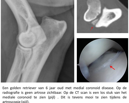
Photo 1 - A 6-year-old golden retriever with medial coronoid disease. No osteoarthritis is visible on the X-ray. The CT scan shows a loose piece of the medial coronoid (arrow). This is also clearly visible during arthroscopy (arrow).
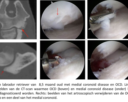
Photo 2 - An 8.5-month-old golden retriever with medial coronoid disease and OCD. Left: images from the CT scan used to diagnose OCD (top) and medial coronoid disease (bottom). Right: images of the arthroscopic removal of the OCD flap and part of the medial coronoid.
Orsami
Universiteit Gent - Faculteit Diergeneeskunde
Salisburylaan 1339820 Merelbeke (Oost-Vlaanderen)België
info.khd@ugent.be+32 9 264 77 00 (kleine huisdieren)+32 9 264 76 18 (grote huisdieren)
Opening hours
| Mo: | 8 - 12h | 12 - 16h |
| Tu: | 8 - 12h | 12 - 16h |
| We: | 8 - 12h | 12 - 16h |
| Th: | 8 - 12h | 12 - 16h |
| Fr: | 8 - 12h | 12 - 16h |
| Sa: | Closed | Closed |
| Su: | Closed | Closed |


